June 12, 2008 — When the world's best sprinters step up to the mark at the Beijing Summer Olympics in August, they will be moments away from subjecting their leg muscles to thousands of pounds of force as they strive to be first across the finish line 10 seconds later. By and large, their leg muscles will handle the strain well, but inevitably one or more of these elite runners, despite intense conditioning, will suffer a hamstring pull.
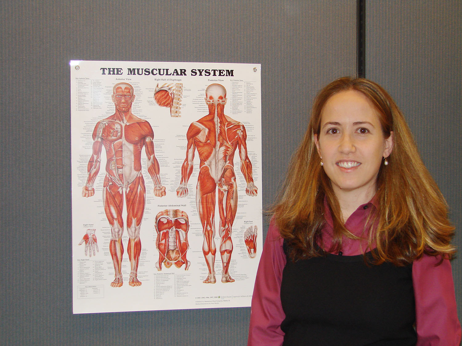
"Of all the muscles that work together when we run quickly, the muscles in the hamstring group are most subject to injury," said Silvia Salinas Blemker, an assistant professor of mechanical and aerospace engineering at the University of Virginia's School of Engineering and Applied Science. "And one particular hamstrings muscle, the biceps femoris long head, is most commonly injured."
Blemker has the expertise in three-dimensional muscle modeling to find out why this muscle is so susceptible to pulls. Collaborating with Darryl Thelen, an associate professor of mechanical engineeering at the University of Wisconsin, she has embarked on a project to identify the points of strain as the biceps femoris moves dynamically and compare it to the other two hamstring muscles. Their research is supported by a four-year, $1.2 million grant from the National Institutes of Health.
The hamstrings run along the back of the thigh and attach on both sides of the knee joint. They are responsible for pulling the foot from the ground with each stride. In the past, researchers treated these muscles like anatomical rubber bands, uniformly elastic along their length. "This simplistic view made it difficult to understand why one muscle is prone to injury while another isn't," she said.
Blemker takes a much more detailed approach, developing finite-element models that incorporate a muscle's intricate internal geometry. But, in order to study injury, Blemker's models need to be analyzed during realistic sprinting movements, which is why Thelen makes an ideal partner. Thelen has developed a model of the whole-body dynamics of sprinting. By combining a finite-element model of the hamstrings with the framework provided by Thelen, Blemker will be able to predict how the muscle will behave in the course of real movement.
Blemker and Thelen face a number of challenges. The first is to merge tissue-level, finite-element models of muscle with whole-body level dynamic models of movement. They will then have to validate their new model by comparing predictions with MRI-imaging techniques that measure muscle strain distribution. "Ultimately, we hope to learn how the internal structure of muscle changes when it is injured, which will help us suggest more effective rehabilitation strategies," she said.
As director of the U.Va. Multiscale Muscle Mechanics Laboratory, Blemker is also developing computational models that connect the properties of individual muscle fibers and the extracellular matrix that binds them together with the properties of the muscle as a whole. This line of research will help us understand how muscles are affected by aging and diseases such as cerebral palsy and muscular dystrophy. She received a Fund for Excellence in Science and Technology Distinguished Young Investigator grant from the University to pursue this work.
Blemker is also collaborating with Dr. Fred Epstein from U. Va.'s radiology department to develop a variety of imaging techniques to characterize the structure and behavior of muscle in living tissue.
Blemker's pioneering work has attracted a number of graduate students, who play a vital role in moving her research projects forward. Michael Rehorn is contributing to the three-dimensional muscle modeling, Bahar Sharafi is working on the fiber and extracellular matrix model, and Niccolo Fiorentino is helping develop imaging techniques to validate the models.
Blemker's work straddles several fields. She has appointments in biomedical engineering and orthopedic surgery as well as mechanical and aerospace engineering, but muscles have always been her focus. "I've been fascinated by the fact that muscles, which are so strong, are so easily injured," she said. "Now I am finding out why."
Media Contact
Article Information
June 12, 2008
/content/engineering-professor-seeks-understand-mechanisms-behind-hamstring-pulls

