Professor George Bloom, an expert in biology, cell biology and neuroscience, said the new tool will facilitate his lab’s research into how subcellular structures within neurons change over the progression of Alzheimer’s disease.
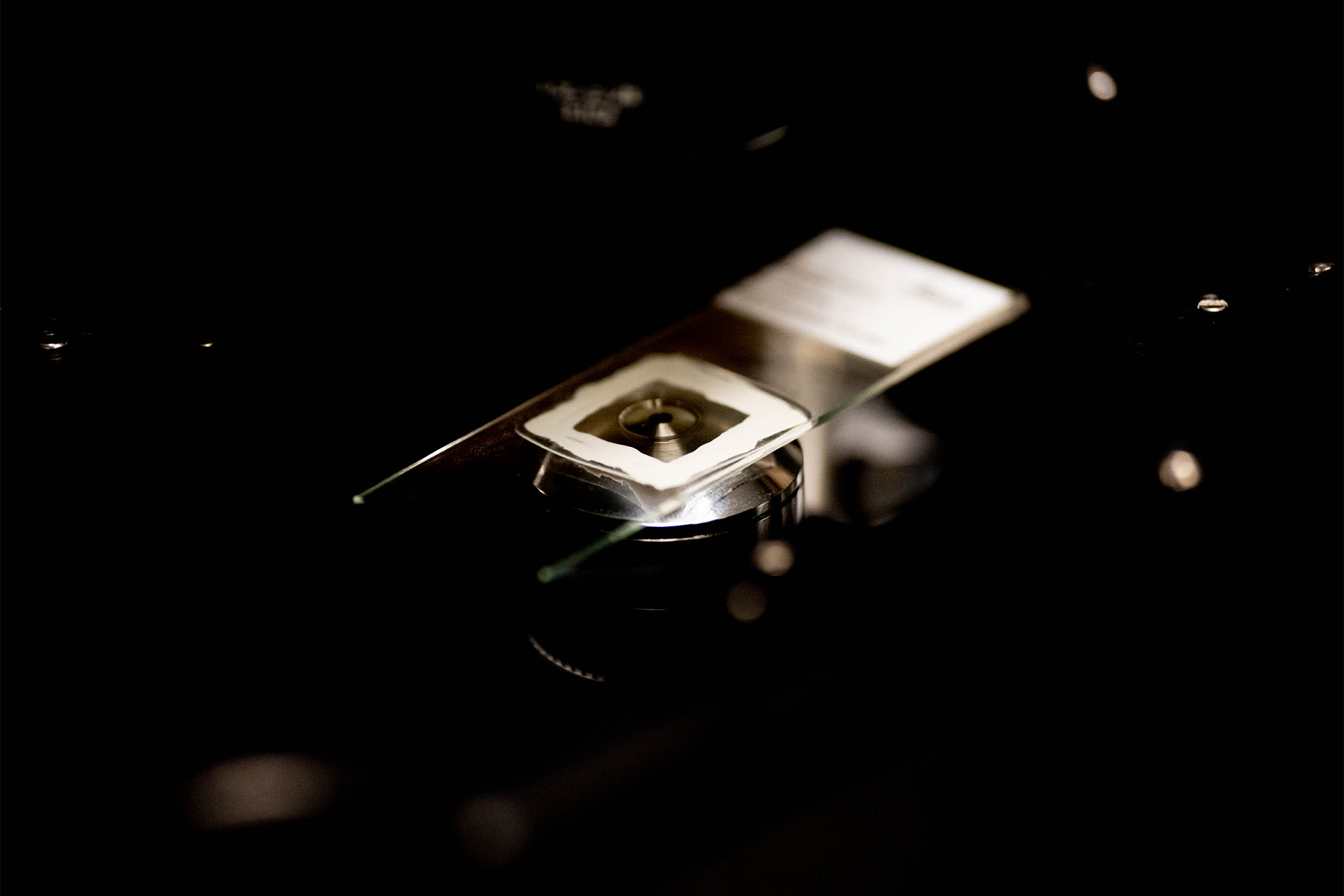
“One of many interesting features of Alzheimer’s disease neurons is that the normally smooth ovoid appearance of their nuclei becomes shriveled and prune-like,” Bloom said. “One of my Ph.D. students, Victoria Sun, discovered what probably causes this phenomenon, and the Leica rep took some beautiful super-resolution images of such a nucleus in a cultured mouse neuron that Victoria treated to become Alzheimer’s disease-like.”
In a demo of the Leica microscope, Alzheimer’s researchers captured a series of “optical sections” of a nucleus at different focus depths. The image stack was then rendered into a rotating, 3-D-like movie.
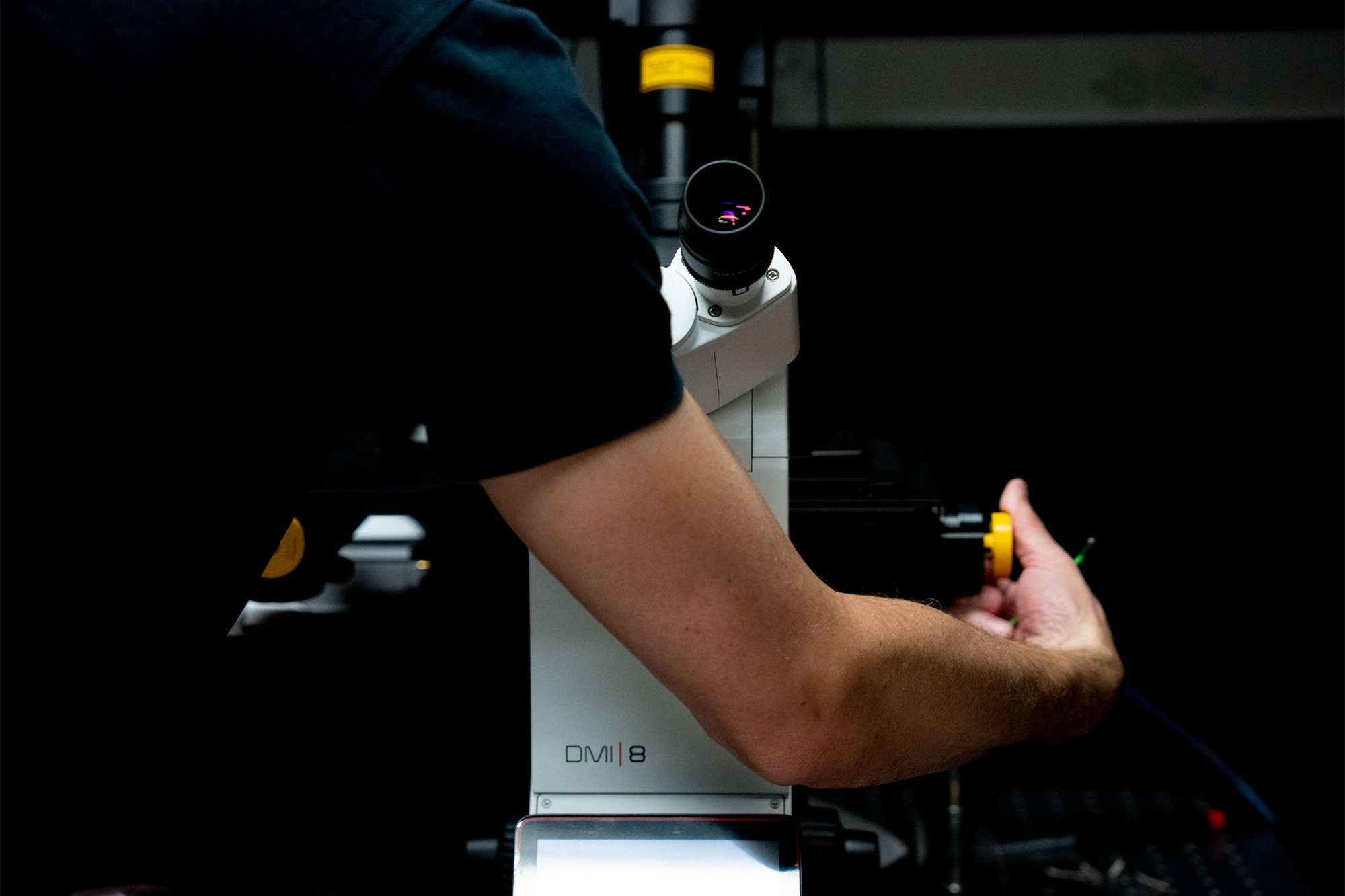
The new system, the latest addition to the W.M. Keck Center for Cellular Imaging, was installed at the end of September and now allows users to see down to 20- to 30-nanometer resolution. To put that into perspective, a sheet of paper is 100,000 nanometers thick.
What’s more, the microscope can produce both two- and three-dimensional images of live cells at between 30- and 70-nanometer resolution.
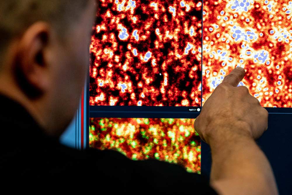
That might mean the difference in a researcher knowing if a protein was even expressed in a biological sample, said Keck Center director Ammasi Periasamy, a professor of biology and biomedical engineering.
“Now they see it; before, they couldn’t,” said Periasamy, who applied for the NIH Office of the Director grant in association with about a dozen UVA researchers who will be using the microscope.