Photos: A Look Inside UVA’s MRI Hub

July 13, 2021 By Caroline Newman, news@virginia.edu Caroline Newman, news@virginia.edu

On a lower floor of the University of Virginia Medical Center, three rooms hum with magnetic resonance imaging scanners so large that they must be lowered in through deconstructed walls or ceilings, secured in place to serve a churn of patients with a wide variety of concerns.

Outside the rooms – which must be entered with care, and without metal, due to the powerful magnetic fields the scanners use – technicians carefully tend to the various monitors tracking patients’ scans, then consult with doctors and nurses who stop by to check in. On any given day they might do several scans of patients’ brains or spines, conduct scans to precisely locate tumors for treatment, or assess the function and structure of the cardiovascular system.

Related Story

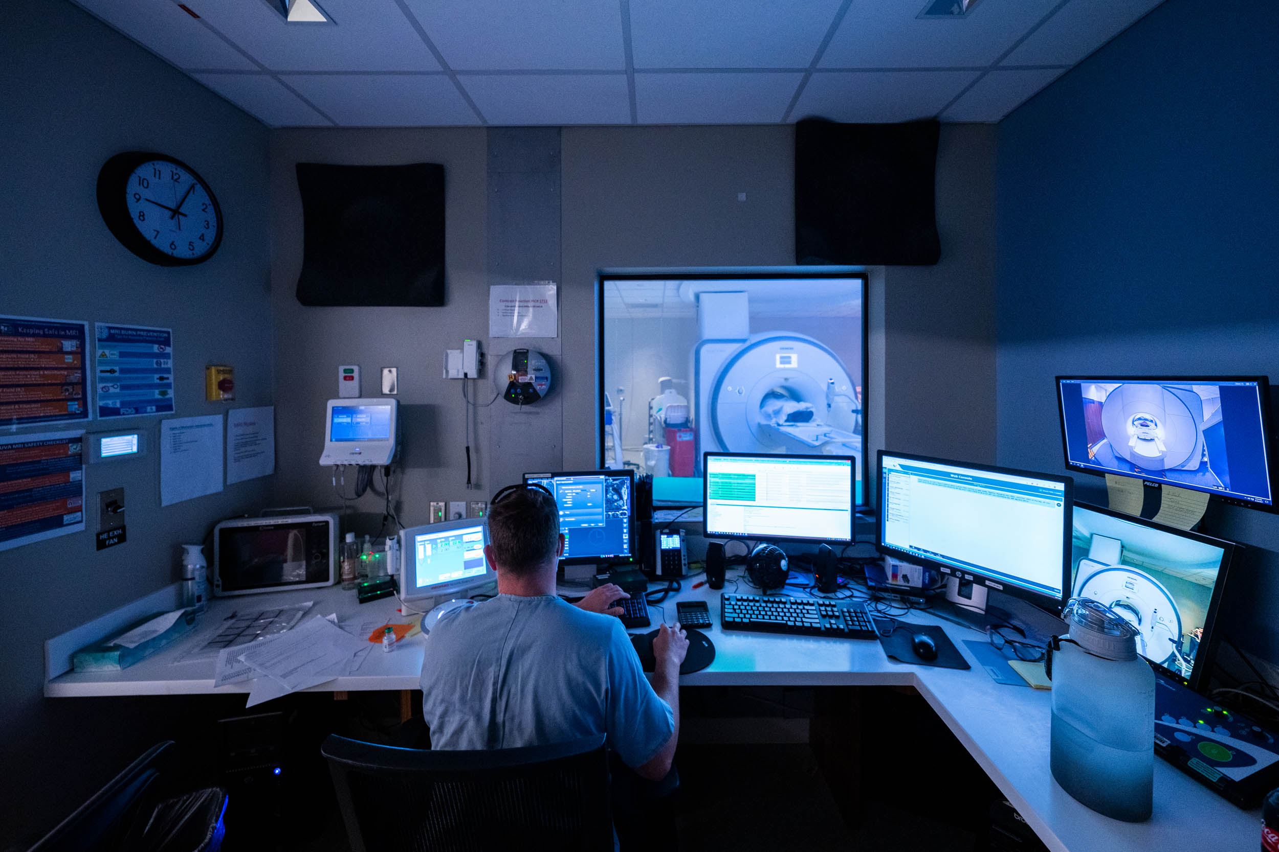
Lead MRI tech Thomas Huerta says that the variety of the work is its own reward; it means that he and his team can touch almost every corner of the UVA and Charlottesville community, and well beyond.

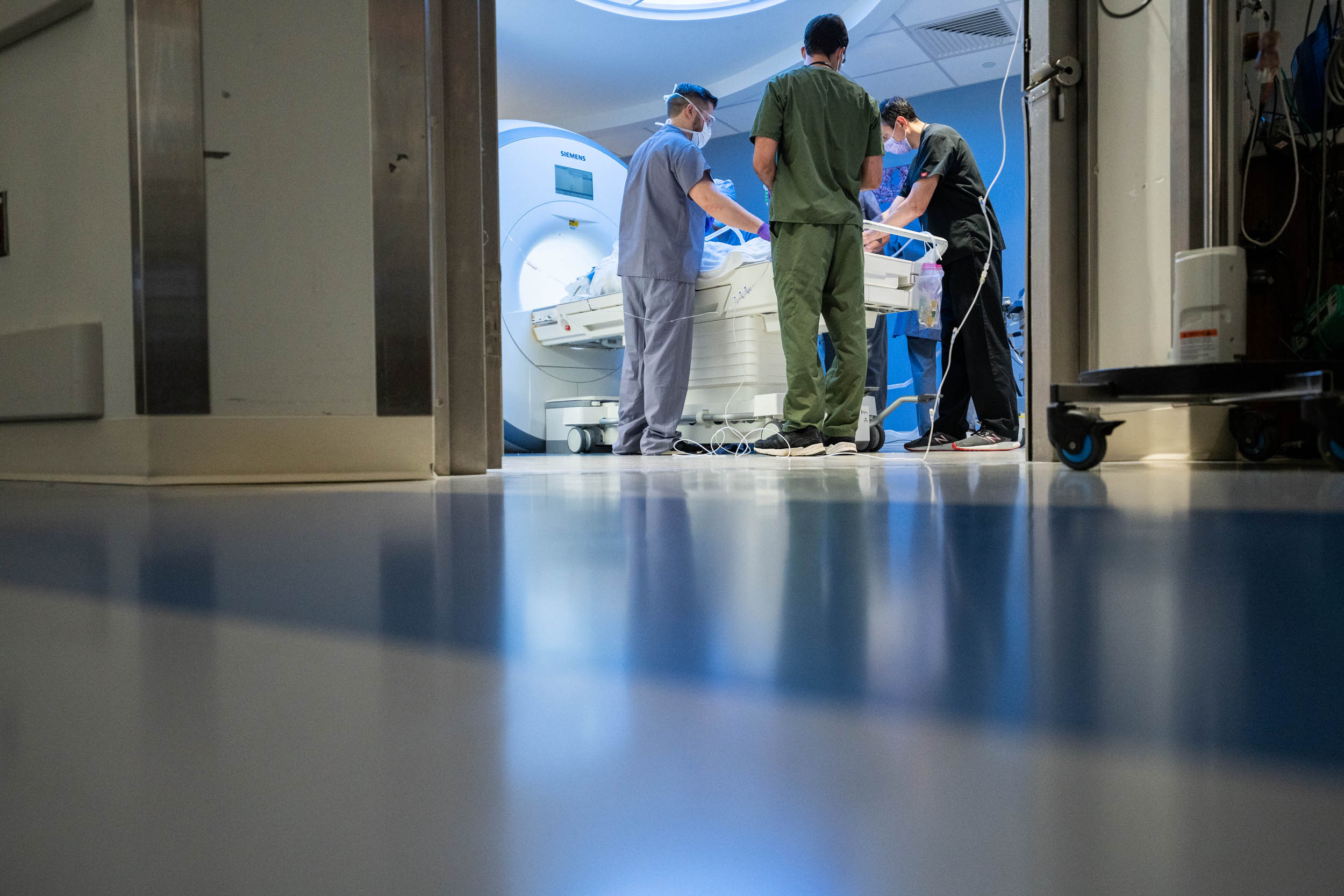
“We do so many different types of MRI here at UVA, and treat kids as young as 4 years old all the way through adulthood,” he said.

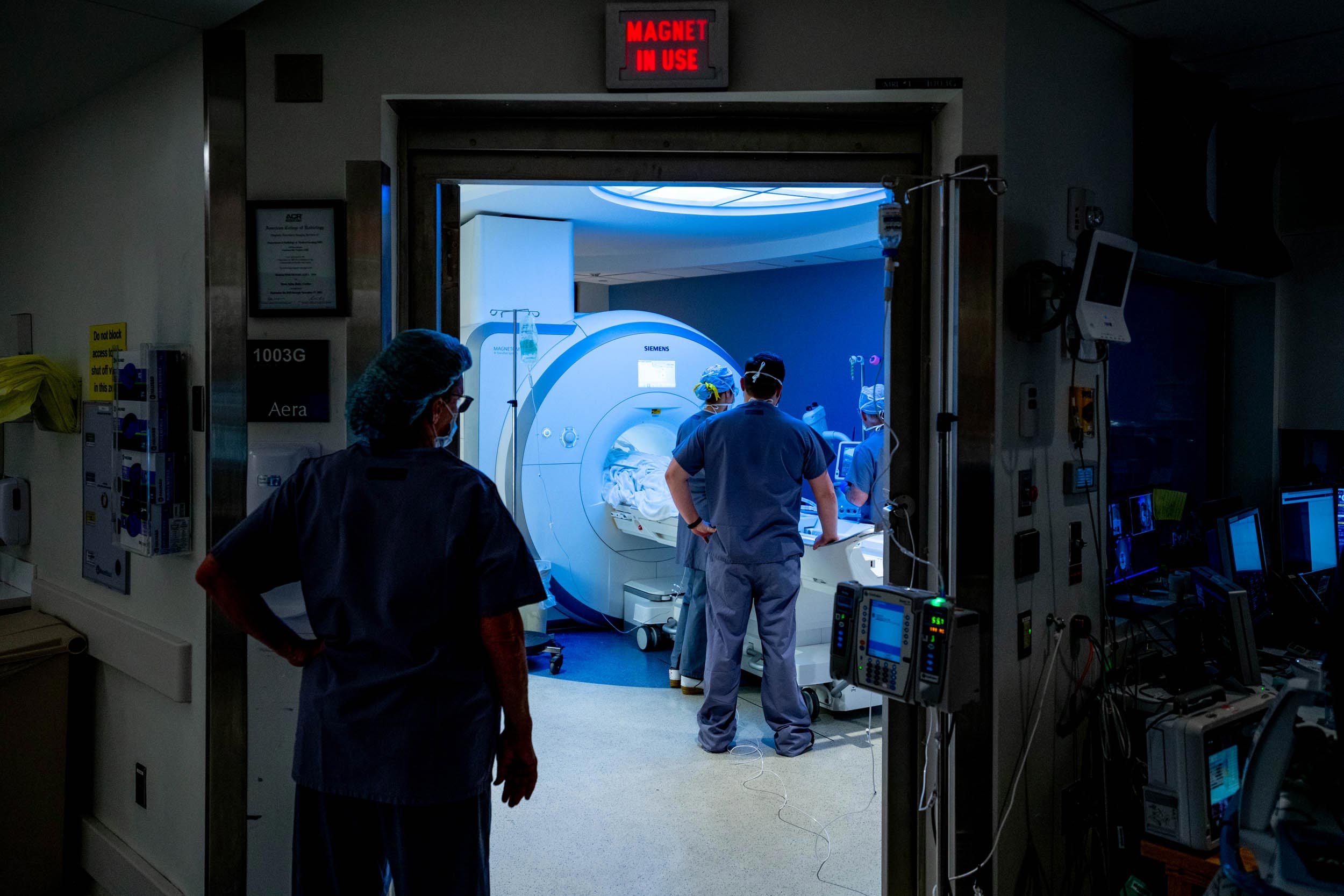
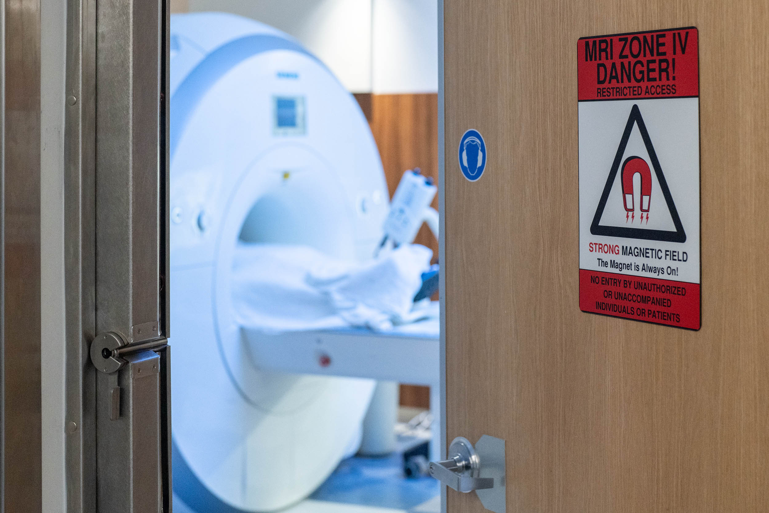
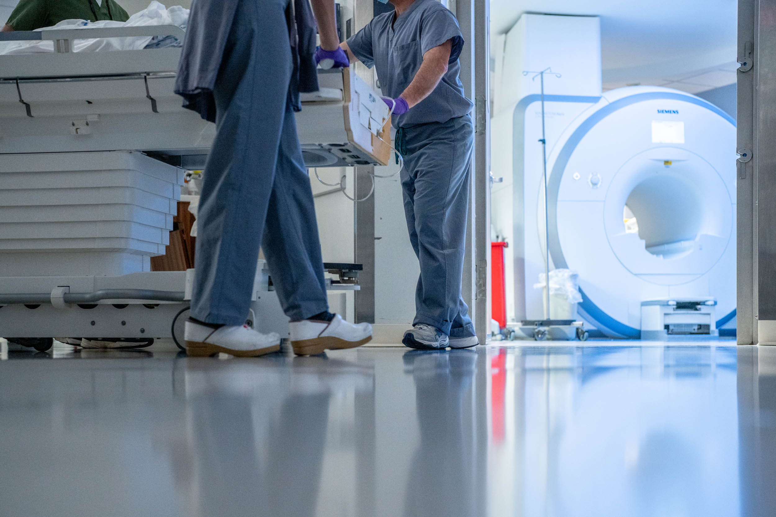
MRI scanners use radio frequency waves to create digital images of the inside of the body. Unlike X-rays, the MRI scans show soft tissue and can be used to examine any part of the body, from head to toe. The scans help doctors diagnose various conditions, look for internal injuries, or see if a medication or treatment is working for diseases like cancer. UVA has three MRI scanners at its main hospital, with additional research scanners housed at Fontaine Research Park. 

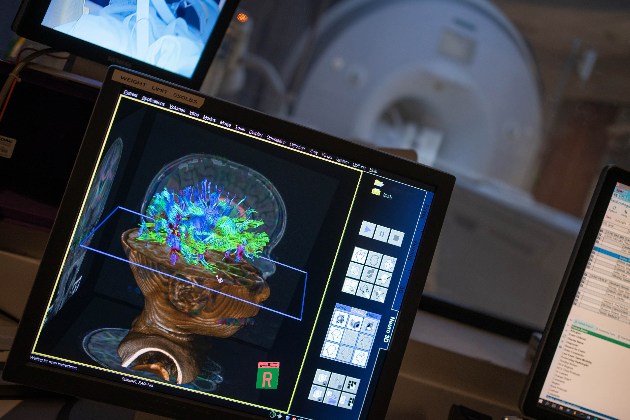
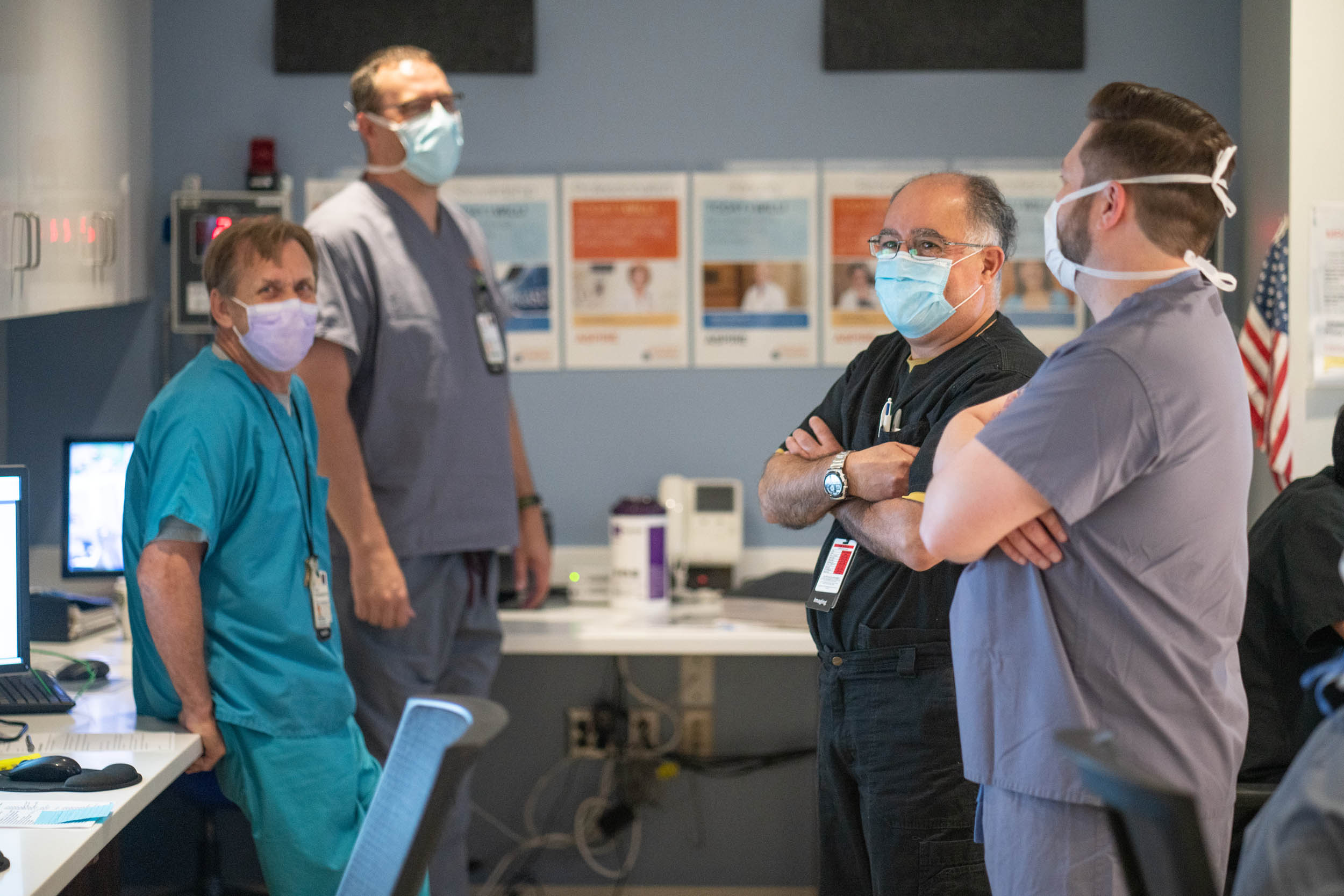
Huerta said UVA’s team of MRI technicians came to the job via many different career and education pathways. He was first introduced to MRI technology in the military, when he trained as a U.S. Army X-ray technician after serving in the airborne infantry. While working at Fort Bragg, North Carolina, he saw an MRI for the first time and was immediately intrigued; you could see so much more than he was used to seeing on X-rays.

“I said right then I was going to do that one day,” he recalled. He pursued MRI certification immediately after leaving the Army. MRI technicians are typically trained as X-ray technicians first and then attend a one-year MRI training program, either through a school or through a job training program.

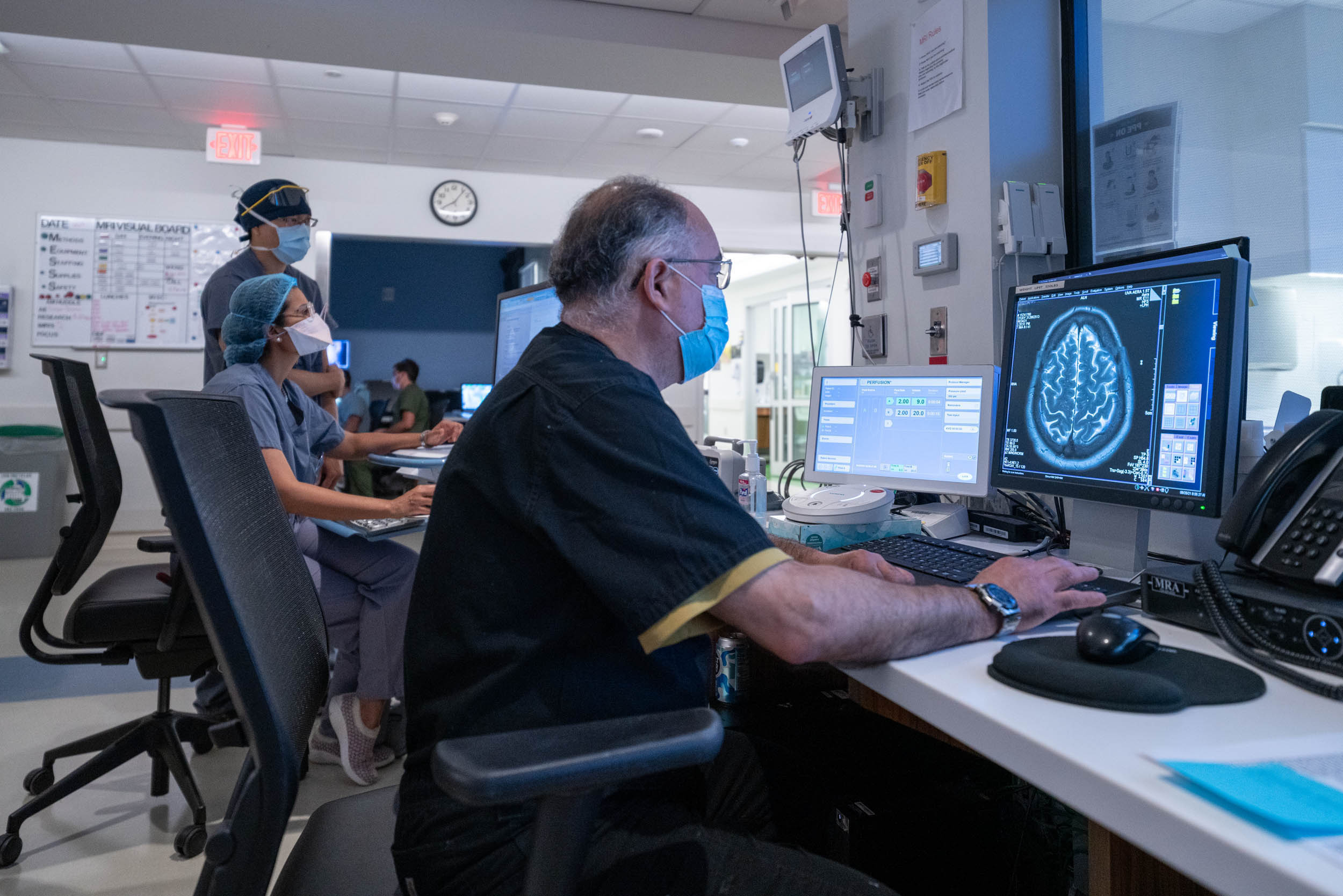
Technicians’ attention to detail is critical because their scans help doctors determine exactly where and how to deliver treatment. The best reward, Huerta said, is hearing that a scan helped doctors uncover the source of a problem or find a new way to treat someone.

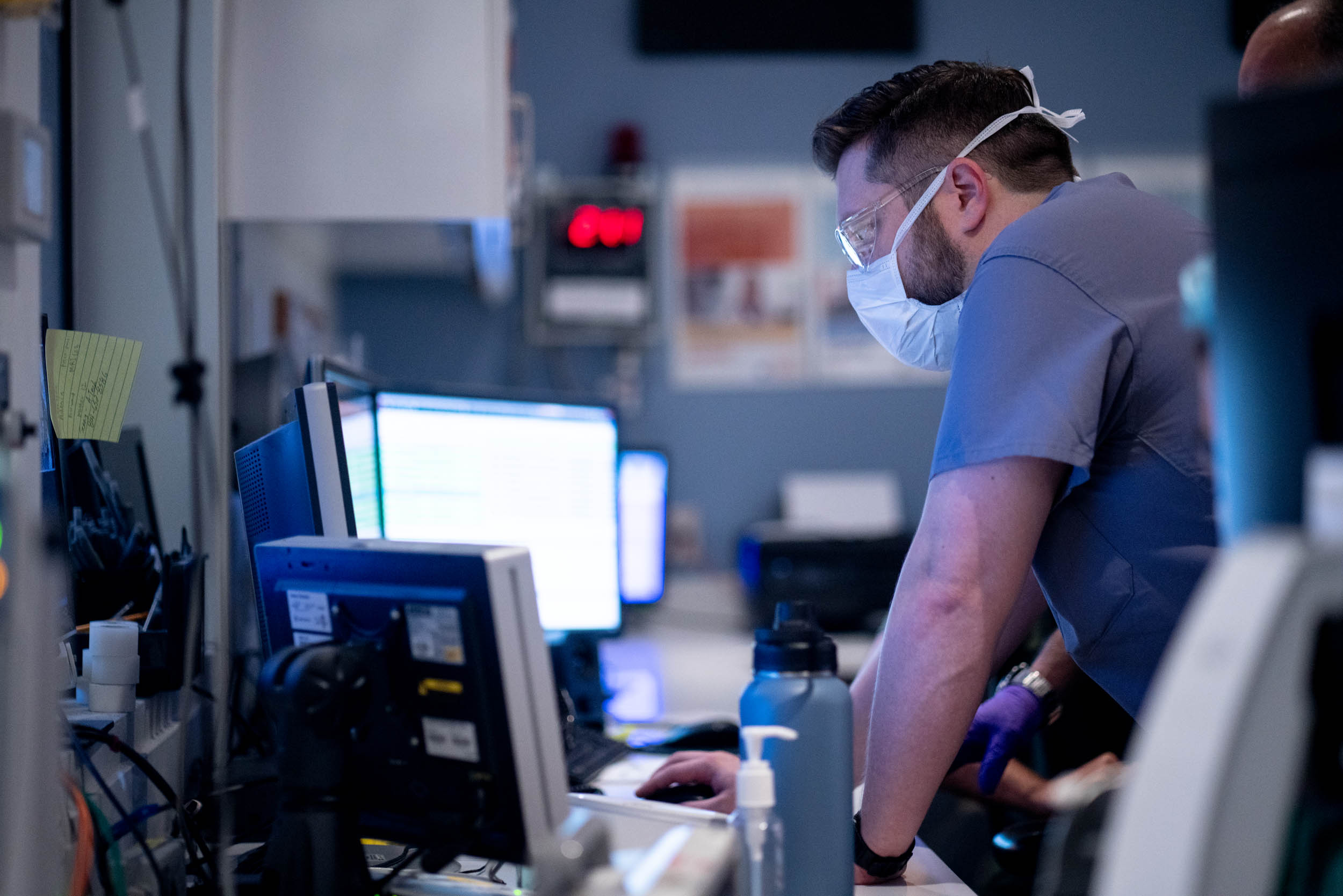
Huerta invited UVA Today to take a look around the MRI unit to see what goes into that work and give readers a window into this common-but-complex diagnostic procedure.

Here’s what it looks like.

Doctor looking at a mri scan of the brain
Thomas Huerta looks through patient scans at a computer station outside of one of the MRI rooms. This particular scan shows an image of the brain; Huerta said brain and spinal scans are among the most frequent MRIs done at UVA.
Person looking at a MRI scan
This scan shows a patient’s heart. MRI scans are a noninvasive way to assess the heart and cardiovascular system.
Computer monitor showing the scan of an MRI on a person
On this particular day, the team was conducting a 3-D brain scan of a pediatric patient who had been suffering from seizures. Doctors will use the scans to seek the cause.
MRI technician sitting at a computer watching and running the MRI machine
A patient is in the scanner, visible through the window, while an MRI technician monitors a bank of computers, positioning and repositioning the scanner to capture every angle needed.
Health professional looking at a computer
Team members took precautions during the pandemic to protect themselves and their patients. They did scan some COVID-19 patients – some with complications from the virus, and others who needed scans for separate ailments.
Health professionals stand in a room talking
Four MRI technicians chat on a quick break as they wait for the next patient.
Healthcare workers preparing a patient for an mri
Team members do what they can to make patients comfortable during procedures, which can last up to 90 minutes. Patients can select their own music playlists, or request podcasts to be piped into the machine.
Door to a MRI open so that we can see the machine.  Door reads MRI Zone Danger!
The scanners’ magnets are always on, meaning that technicians must always be careful to remove any metal before entering the room, and to remind patients and other staff to do the same.
Health care professionals pushing patient into the room with an MRI
Stretchers used in the MRI units are specially made without magnetic materials, so patients can be transferred seamlessly – another way to the team emphasizes both safety and comfort for patients.