Ending a 50-Year Mystery, UVA Reveals How Bacteria Can Move

September 26, 2022
Illustration of hairy, pill shaped bacteria

For 50 years, scientists didn’t know exactly how bacteria moved, but UVA researchers and colleagues using a special microscope solved the mystery. (Illustration by Emily Faith Morgan, University Communications).

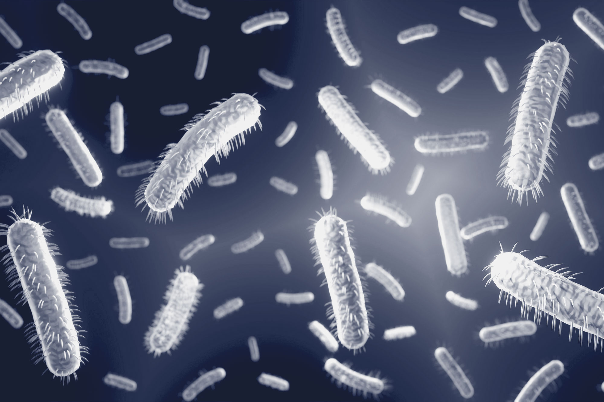
University of Virginia School of Medicine researchers and their collaborators have solved a decades-old mystery about how E. coli and other bacteria are able to move.

Bacteria push themselves forward by coiling long, threadlike appendages into corkscrew shapes that act as makeshift propellers. But how exactly they do this has baffled scientists, because the “propellers” are made of a single protein. 

An international team led by UVA’s Edward H. Egelman, a leader in the field of high-tech cryo-electron microscopy, has cracked the case. The researchers used the advanced microscopy, sometimes called cryo-EM, and advanced computer modeling to reveal what no traditional light microscope could see: the strange structure of these propellers at the level of individual atoms.

“While models have existed for 50 years for how these filaments might form such regular coiled shapes, we have now determined the structure of these filaments in atomic detail,” said Egelman, of UVA’s Department of Biochemistry and Molecular Genetics. “We can show that these models were wrong, and our new understanding will help pave the way for technologies that could be based upon such miniature propellers.”

Blueprints for Bacteria’s ‘Supercoils’

Different bacteria have one or many appendages known as a flagellum, or, in the plural, flagella. A flagellum is made of thousands of subunits, but all these subunits are exactly the same. You might think that such a tail would be straight, or at best a bit flexible, but that would leave the bacteria unable to move. That’s because such shapes can’t generate thrust. It takes a rotating, corkscrew-like propeller to push a bacterium forward. Scientists call the formation of this shape “supercoiling,” and now, after more than five decades, they understand how bacteria do it.

Egelman stands with his hands clasped and looks at the camera
UVA Researcher Edward Egelman said the discovery showed that previous models guessing how bacteria moved about “were wrong.” (Photo by Dan Addison, University Communications).

Using cryo-EM, Egelman and his team found that the protein that makes up the flagellum can exist in 11 different states. It is the precise mixture of these states that causes the corkscrew shape to form.

It has been known that the propeller in bacteria is quite different than similar propellers used by hardy one-celled organisms called archaea. Archaea are found in some of the most extreme environments on Earth, such as in nearly boiling pools of acid, the very bottom of the ocean and in petroleum deposits deep in the ground.

Egelman and colleagues used cryo-EM to examine the flagella of one form of archaea, Saccharolobus islandicus, and found that the protein forming its flagellum exists in 10 different states. While the details were quite different than what the researchers saw in bacteria, the result was the same, with the filaments forming regular corkscrews. They conclude that this is an example of “convergent evolution” – when nature arrives at similar solutions via very different means. This shows that even though bacteria and archaea’s propellers are similar in form and function, the organisms evolved those traits independently.

Daily Report
The latest UVA news, delivered to your inbox.

“As with birds, bats and bees, which have all independently evolved wings for flying, the evolution of bacteria and archaea has converged on a similar solution for swimming in both,” said Egelman, whose prior imaging work saw him inducted into the National Academy of Sciences, one of the highest honors a scientist can receive. “Since these biological structures emerged on Earth billions of years ago, the 50 years that it has taken to understand them may not seem that long.”  

Findings Published

The researchers have published their findings in the scientific journal Cell. The team consisted of Mark A.B. Kreutzberger, Ravi R. Sonani, Junfeng Liu, Sharanya Chatterjee, Fengbin Wang, Amanda L. Sebastian, Priyanka Biswas, Cheryl Ewing, Weili Zheng, Frédéric Poly, Gad Frankel, B.F. Luisi, Chris Calladine, Mart Krupovic, Birgit E. Scharf and Egelman. 

The work was supported by the National Institutes of Health, grants GM122150 and T32 GM080186; U.S. Navy Work Unit Program 6000.RAD1.DA3.A0308; and by a Robert R. Wagner Fellowship. The researchers’ paper does not represent the official policy or position of the Department of the Navy, Department of Defense or the U.S. government.

To keep up with the latest medical research news from UVA, subscribe to the Making of Medicine blog.

Media Contact

Josh Barney

UVA Health