August 16, 2011 — While there is plenty of research to show how the outside of muscles work under strained conditions, researchers are just beginning to get an accurate look beneath the muscle surface and into connective tissues. From this vantage, researchers will be able to isolate the biomechanics that lead to injury, as well as pinpoint and treat genes responsible for hereditary disorders such as Duchenne muscular dystrophy.
Two University of Virginia mechanical engineering graduate students were recently honored for research that will help improve researchers' understanding of muscle injury by advancing biomechanical computer simulation. Bahar Sharafi, a recent Ph.D. graduate, and Nic Fiorentino, a Ph.D. candidate in the Department of Mechanical and Aerospace Engineering, won first and second place, respectively, for the Komor New Investigator Award at the International Society for Biomechanics Computer Simulation. The symposium was held in July in Belgium.
Listen to the UVA Today Radio Show report on this story by Marian Anderfuren:
Sharafi and Fiorentino both work in associate professor Silvia Blemker's Multi-scale Muscle Mechanics Lab in U.Va.'s School of Engineering and Applied Science. Blemker has a joint appointment in departments of Mechanical and Aerospace Engineering and Biomedical Engineering.
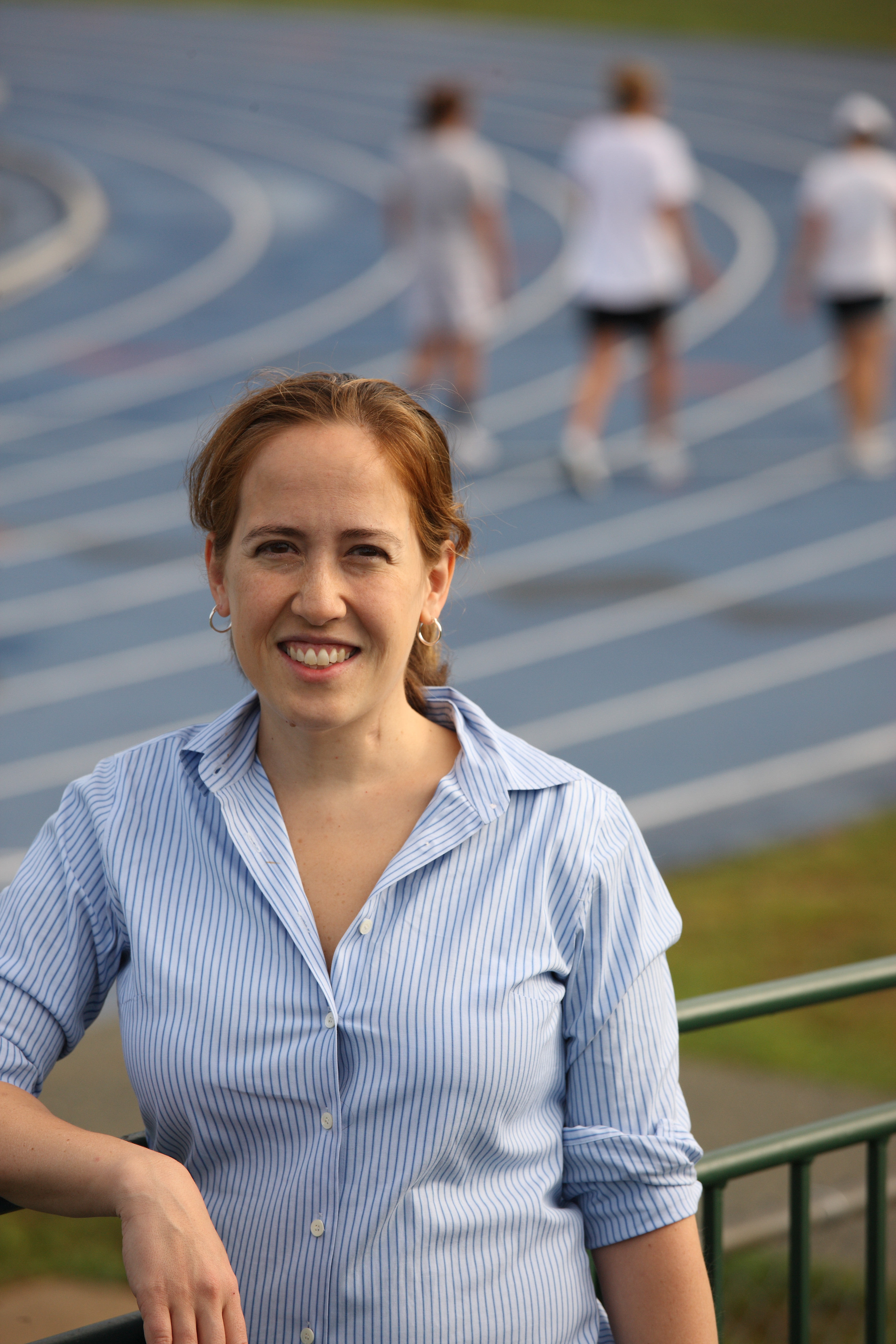
Blemker and her team of researchers create computer models of skeletal muscles to answer questions that will help eventually improve the work of athletic trainers, physical therapists and other medical professionals who treat patients with muscle injuries and disorders.
"Not much is known about how muscles behave in vivo, or within the muscles themselves, and we're building computer models to figure it out," Blemker said. "These models can predict how a muscle is behaving beneath its surface and help us understand why injury occurs."
A key to Blemker's research is discovering how stretch and contraction can injure a complex network of fibers and tendons inside of muscles and connective tissues.
For the recent symposium, Sharafi presented a fiber-scale micromechanical model that looked at the individual fibers and their connections with tendons. The model predicts how fibers deform when muscles stretch. She experimented by imaging muscle fibers in slack and stretch position and measured striation patterns in a range of positions. The image showed how deformation occurs between the two positions.
Fiorentino complemented Sharafi's talk with a presentation about a macro-model he developed to show the entire muscle-tendon unit.
His model shows how muscle tissue behaves during lengthening. He performed activated, dynamic experiments using magnetic resonance imaging – or MRI – to show how strains occur during knee exercise. The exercise experiment required subjects to exert a lesser degree of force than what would lead to injury, but still represent an injurious activity. The results of the simulated MRI experiment showed a strong correlation with the striation produced in Fiorentino's computer model.
Blemker believes one reason Sharafi and Fiorentino outshone other presenters during the recent symposium was that they successfully used experiments to validate their models.
"In our work, it can be difficult to perform the complex experiments required to validate models," Blemker said. "They successfully did so and made a powerful scientific case."
One main pursuit of the lab's research is to use large-scale muscle models to identify features of muscles that make them more susceptible to injury. Through MRI experiments, they can further determine why some people are more predisposed to injury than others are. In the case of training to prevent injury, Blemker and her researchers are beginning collaborations with the U.Va. track team to answer some of those questions.
On the micro-level, the researchers are most excited about better understanding the origins and treatments of Duchenne muscular dystrophy. Researchers believe the condition occurs from either the deletion or mutation of a gene in the muscles between fibers and in the connective tissue surrounding fibers. The muscles of people with the disease are highly susceptible to injury from routine activity and exercise. Children with the disorder usually do not live past their 20s because of their weakened hearts and lungs.
"We need to understand how this protein protects muscles from injury and apply that understanding to treatment strategies for replacing or treating the gene," Blemker said.
Bahar will start a post-doctoral research scientist position at Northwestern University this fall. Fiorentino plans to finish his Ph.D. in May.
Two University of Virginia mechanical engineering graduate students were recently honored for research that will help improve researchers' understanding of muscle injury by advancing biomechanical computer simulation. Bahar Sharafi, a recent Ph.D. graduate, and Nic Fiorentino, a Ph.D. candidate in the Department of Mechanical and Aerospace Engineering, won first and second place, respectively, for the Komor New Investigator Award at the International Society for Biomechanics Computer Simulation. The symposium was held in July in Belgium.
Listen to the UVA Today Radio Show report on this story by Marian Anderfuren:
Sharafi and Fiorentino both work in associate professor Silvia Blemker's Multi-scale Muscle Mechanics Lab in U.Va.'s School of Engineering and Applied Science. Blemker has a joint appointment in departments of Mechanical and Aerospace Engineering and Biomedical Engineering.
Blemker and her team of researchers create computer models of skeletal muscles to answer questions that will help eventually improve the work of athletic trainers, physical therapists and other medical professionals who treat patients with muscle injuries and disorders.
"Not much is known about how muscles behave in vivo, or within the muscles themselves, and we're building computer models to figure it out," Blemker said. "These models can predict how a muscle is behaving beneath its surface and help us understand why injury occurs."
A key to Blemker's research is discovering how stretch and contraction can injure a complex network of fibers and tendons inside of muscles and connective tissues.
For the recent symposium, Sharafi presented a fiber-scale micromechanical model that looked at the individual fibers and their connections with tendons. The model predicts how fibers deform when muscles stretch. She experimented by imaging muscle fibers in slack and stretch position and measured striation patterns in a range of positions. The image showed how deformation occurs between the two positions.
Fiorentino complemented Sharafi's talk with a presentation about a macro-model he developed to show the entire muscle-tendon unit.
His model shows how muscle tissue behaves during lengthening. He performed activated, dynamic experiments using magnetic resonance imaging – or MRI – to show how strains occur during knee exercise. The exercise experiment required subjects to exert a lesser degree of force than what would lead to injury, but still represent an injurious activity. The results of the simulated MRI experiment showed a strong correlation with the striation produced in Fiorentino's computer model.
Blemker believes one reason Sharafi and Fiorentino outshone other presenters during the recent symposium was that they successfully used experiments to validate their models.
"In our work, it can be difficult to perform the complex experiments required to validate models," Blemker said. "They successfully did so and made a powerful scientific case."
One main pursuit of the lab's research is to use large-scale muscle models to identify features of muscles that make them more susceptible to injury. Through MRI experiments, they can further determine why some people are more predisposed to injury than others are. In the case of training to prevent injury, Blemker and her researchers are beginning collaborations with the U.Va. track team to answer some of those questions.
On the micro-level, the researchers are most excited about better understanding the origins and treatments of Duchenne muscular dystrophy. Researchers believe the condition occurs from either the deletion or mutation of a gene in the muscles between fibers and in the connective tissue surrounding fibers. The muscles of people with the disease are highly susceptible to injury from routine activity and exercise. Children with the disorder usually do not live past their 20s because of their weakened hearts and lungs.
"We need to understand how this protein protects muscles from injury and apply that understanding to treatment strategies for replacing or treating the gene," Blemker said.
Bahar will start a post-doctoral research scientist position at Northwestern University this fall. Fiorentino plans to finish his Ph.D. in May.
— By Zak Richards
Media Contact
Article Information
August 16, 2011
/content/looking-inside-muscles-uva-students-honored-advancing-biomechanical-computer-simulation