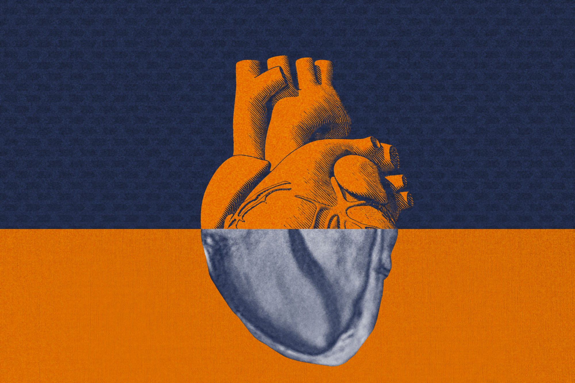
A new artificial-intelligence technology for heart imaging can potentially improve care for patients, allowing doctors to examine their hearts for scar tissue while eliminating the need for contrast injections required for traditional cardiovascular magnetic resonance imaging.
A team of researchers who developed the technology, including doctors at UVA Health, reports the success of the approach in a new article in the scientific journal Circulation. The team compared its AI approach, known as virtual native enhancement, with contrast-enhanced cardiovascular magnetic resonance scans now used to monitor hypertrophic cardiomyopathy, the most common genetic heart condition. The researchers found that virtual native enhancement produced higher-quality images and better captured evidence of scar in the heart, all without the need for injecting the standard contrast agent required for cardiovascular magnetic resonance scans.
“This is a potentially important advance, especially if it can be expanded to other patient groups,” said researcher Dr. Christopher Kramer, the chief of the Division of Cardiovascular Medicine at UVA Health, Virginia’s only designated Center of Excellence by the Hypertrophic Cardiomyopathy Association. “Being able to identify scar in the heart, an important contributor to progression to heart failure and sudden cardiac death, without contrast, would be highly significant. Cardiovascular magnetic resonance scans would be done without contrast, saving cost and any risk, albeit low, from the contrast agent.”
Imaging Hypertrophic Cardiomyopathy
Hypertrophic cardiomyopathy is the most common inheritable heart disease, and the most common cause of sudden cardiac death in young athletes. It causes the heart muscle to thicken and stiffen, reducing its ability to pump blood and requiring close monitoring by doctors.
The new virtual native enhancement technology will allow doctors to image the heart more often and more quickly, the researchers say. It also may help doctors detect subtle changes in the heart earlier, though more testing is needed to confirm that.
The technology also would benefit patients who are allergic to the contrast agent injected for cardiovascular magnetic resonance scans, as well as patients with severely failing kidneys, a group that avoids the use of the agent.
The new approach works by using artificial intelligence to enhance “T1-maps” of the heart tissue created by magnetic resonance imaging. These maps are combined with enhanced MRI “cines,” which are like movies of moving tissue – in this case, the beating heart. Overlaying the two types of images creates the artificial virtual native enhancement image.
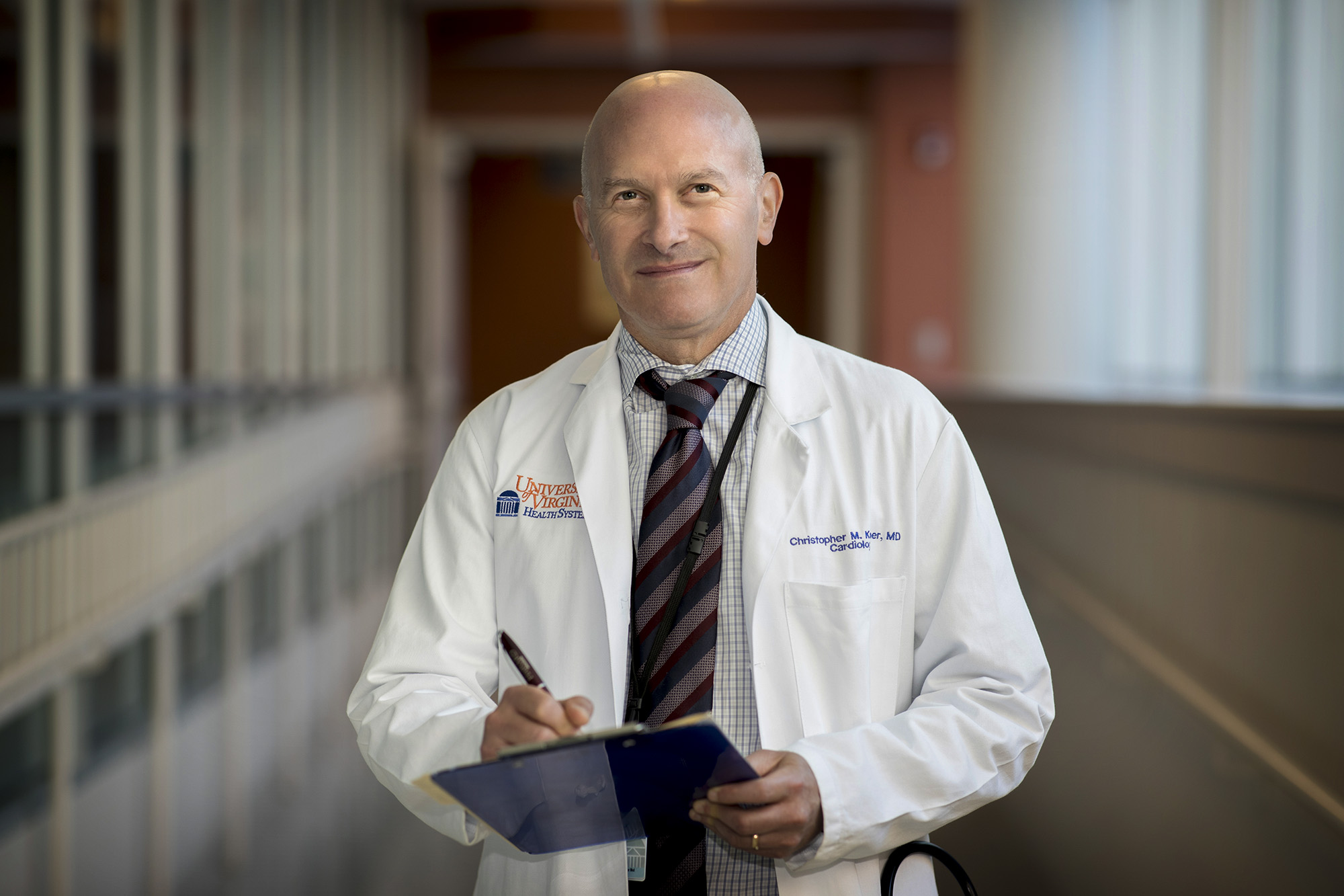
Dr. Christopher Kramer is the chief of the Division of Cardiovascular Medicine at UVA Health. (Photo by Dan Addison, University Communications)
Based on these inputs, the technology can produce something virtually identical to the traditional contrast-enhanced cardiovascular magnetic resonance heart scans doctors are accustomed to reading – only better, the researchers conclude. “Avoiding the use of contrast and improving image quality in [cardiovascular magnetic resonance] would only help both patients and physicians down the line,” Kramer said.
While the new research examined virtual native enhancement’s potential in patients with hypertrophic cardiomyopathy, the technology’s creators envision it being used for many other heart conditions as well.
“While currently validated in the [hypertrophic cardiomyopathy] population, there is a clear pathway to extend the technology to a wider range of myocardial pathologies,” they write. “[Virtual native enhancement] has enormous potential to significantly improve clinical practice, reduce scan time and costs, and expand the reach of [cardiovascular magnetic resonance] in the near future.”
About the Research
The research team consisted of Qiang Zhang, Matthew K. Burrage, Elena Lukaschuk, Mayooran Shanmuganathan, Iulia A. Popescu, Chrysovalantou Nikolaidou, Rebecca Mills, Konrad Werys, Evan Hann, Ahmet Barutcu, Suleyman D. Polat, HCMR investigators, Michael Salerno, Michael Jerosch-Herold, Raymond Y. Kwong, Hugh C. Watkins, Christopher M. Kramer, Stefan Neubauer, Vanessa M. Ferreira and Stefan K. Piechnik.
Kramer has no financial interests in the research, but some of his collaborators are seeking a patent related to the imaging approach. A full list of disclosures is included in the paper.
The research was made possible by work funded by the British Heart Foundation, grant PG/15/71/31731; the National Institutes of Health’s National Heart, Lung and Blood Institute, grant U01HL117006-01A1; the John Fell Oxford University Press Research Fund; and the Oxford BHF Centre of Research Excellence, grant RE/18/3/34214. The research was also supported by British Heart Foundation Clinical Research Training Fellowship FS/19/65/34692, National Institute for Health Research (NIHR) Oxford Biomedical Research Centre at The Oxford University Hospitals NHS Foundation Trust, and the National Institutes of Health.
To keep up with the latest medical research news from UVA, subscribe to the Making of Medicine blog.
Media Contact
Article Information
August 31, 2021
/content/new-artificial-intelligence-technology-poised-transform-heart-imaging