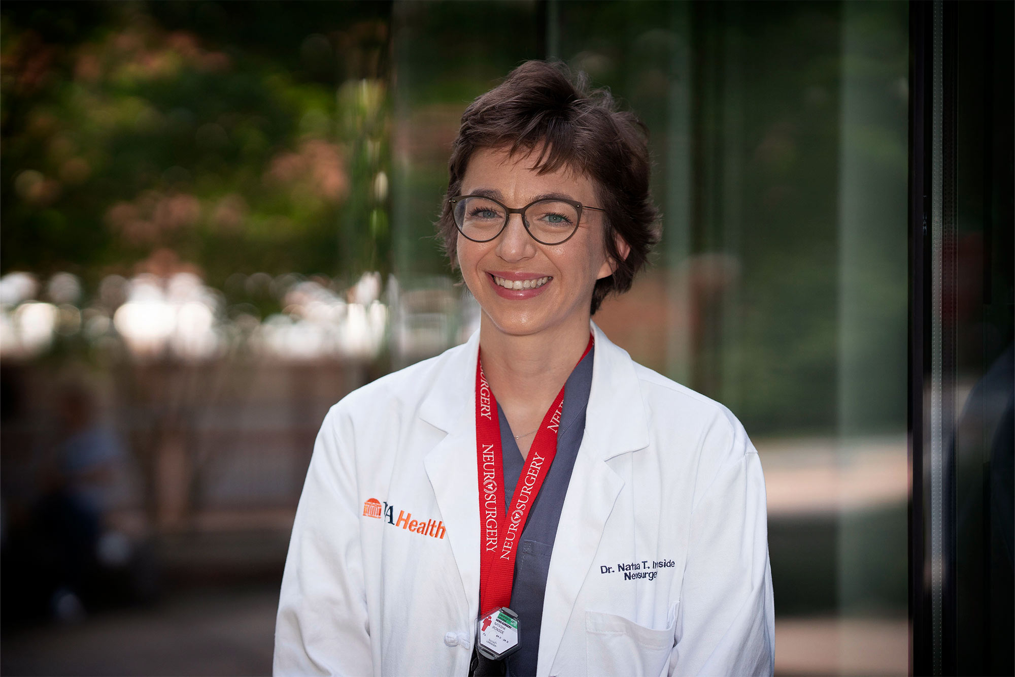
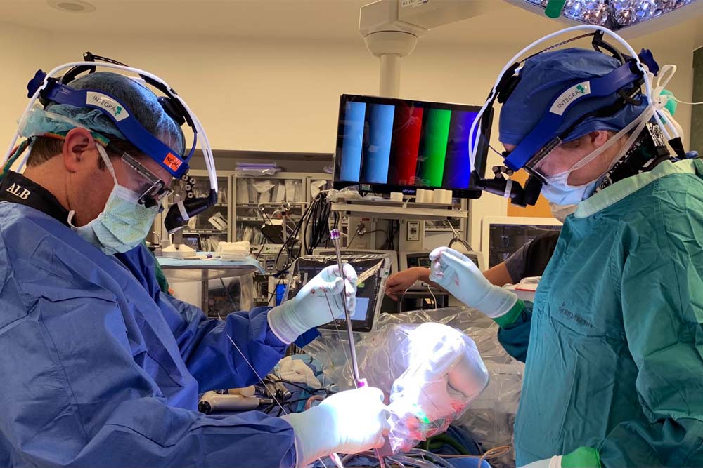
In Dr. Natasha Ironside’s world, there are a number of brains at work. There’s her brain, of course; she’s a University of Virginia neurosurgery resident who’s always thinking about how to improve stroke outcomes in the brains of her patients. And then there’s the artificially intelligent “brain” she created to help with the problem.
While a research student at Columbia University, she taught herself how to code, and the subsequent artificial intelligence tool she programmed was able to quickly and accurately quantify the extent of brain bleeds, or hematomas, and swelling (edema) captured on CT scans.
“During this period, I realized that it was incredibly challenging and time-consuming to manually measure both the hemorrhage and surrounding edema volumes on CT scans,” Ironside said. “We needed a more efficient and standardized way of measuring.”
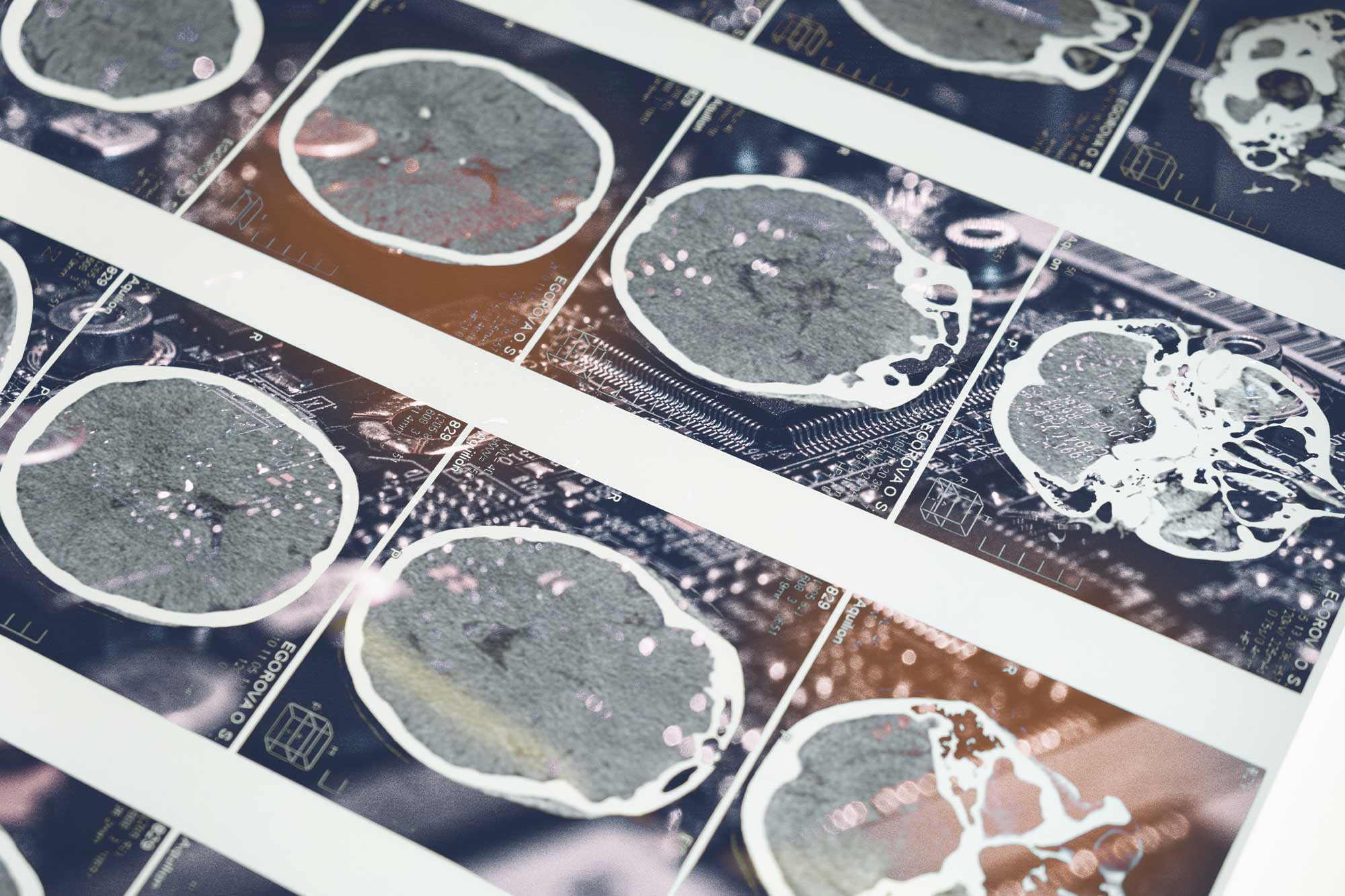
At Columbia, her AI performed as well or better than clinicians working with existing programs to understand the extent of intracerebral hemorrhage – or bleeding into the brain tissue. That’s the second-leading cause of stroke, so having the quickest, most accurate understanding of the parameters of an injury can help doctors like her save lives.
“We showed that the AI tool can replicate human analyses and semi-automated machine-based assessments of the images, with high accuracy,” Ironside said. “We also demonstrated that the AI program performs analyses at a much faster rate.”
She published the findings in the journal Stroke in 2020.
So how fast is fast? It takes clinicians about 25 minutes on average to quantify both hematoma and edema on a CT scan. Her AI tool can compute these volumes in approximately 30 seconds.
“This is especially important when dealing with a large number of CT scans in a clinical setting, when fast decision-making is necessary to implement treatments in a timely manner,” she said.
With her AI having performed successfully in the preliminary study, she devised a more definitive study by collaborating with several other university medical systems, including UVA Health. That study is now wrapping up. The study includes six “blinded” expert raters – neuroradiologists, neurosurgeons and neurologists – looking at CT scans of patients with brain bleeds.
The goal of the further study is to indisputably establish that measurements from the AI program are equivalent to those done by human experts.
Among other functions, the new study clarifies the parameters of what constitutes an “equivalent” measure, and it minimizes the potential biases among raters. The raters and investigators are prevented from seeing the results, and the study is being monitored by an independent monitoring board.
Currently, no standardized methods for testing the safety and reliability of AI programs in health care exist. Ironside hopes her study, which she has dubbed QUANTUM (Quantification of Hematoma and Perihematomal Edema Volumes in Intracerebral Hemorrhage), will set the standard going forward.
UVA researchers participating in the study are Karen Johnston, Sugoto Mukherjee, Min S. Park, James Patrie, Tanvir Rizvi, Andrew Southerland and Bradford Worrall.
Participating from other universities are Ching-Jen Chen of the University of Texas Health Science Center at Houston, Jan Claassen and E. Sander Connolly of Columbia University, Dale Ding of the University of Louisville, and Pascal Jabbour of Thomas Jefferson University.
Chen, a graduate of the UVA Department of Neurosurgery who is now a new faculty member at the University of Texas, said he’s excited to see Ironside’s technology tested on such a wide scale.
“Rigorous validation of the AI is critical in providing a solid foundation for its future and wider applications in stroke care and medicine,” Chen said.
Results of the QUANTUM study are expected at the end of this year.
Media Contact
Communications Manager School of Engineering and Applied Science
williamson@virginia.edu (434) 924-1321
Article Information
November 25, 2024