


There’s a good chance that if you’ve ever needed a magnetic resonance imaging scan – an MRI – you benefited from one of Craig Meyer’s inventions, and that may have made a world of difference in your treatment and prognosis.
In the early 1980s, Meyer saw a story in a magazine about the way this seemingly futuristic machine could safely peer into the body using physics, math and magnets. He was immediately intrigued; and because the technology hit all his goal-oriented buttons of doing good in the world using applied mathematics and science, he jumped at the chance to study the first-ever commercial MRI machine that arrived at Stanford University shortly after he did.
Within just a few short years of Meyer’s work at Stanford, the inquisitive Midwesterner devised a system that sped up image-making and analysis, further paving the way for the huge machines to have realistic applications in clinical settings. This early work resulted in his first patent in 1991.
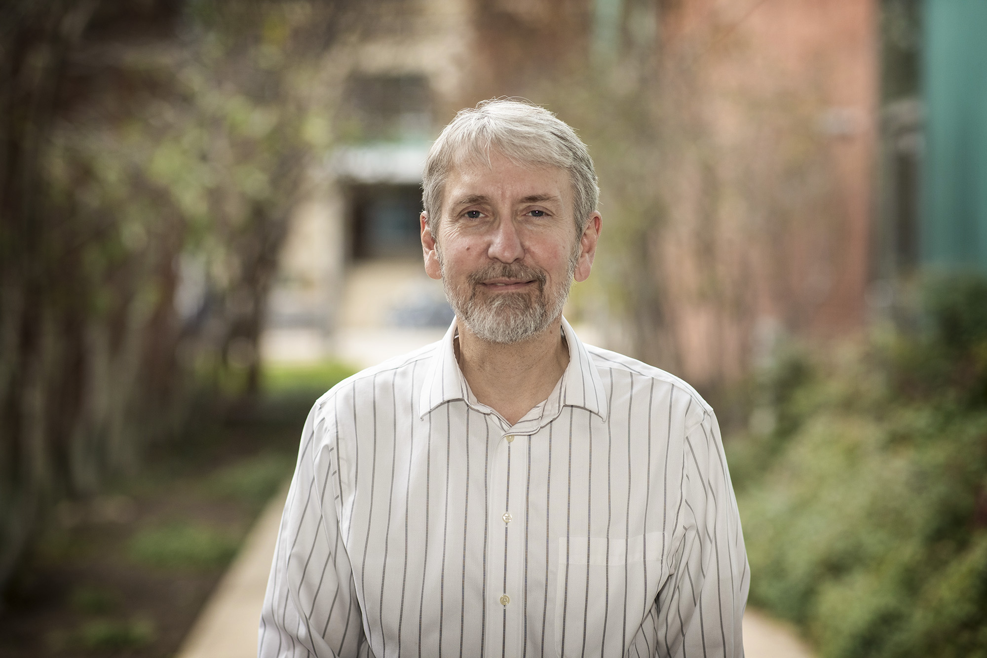
Today, more than 36 patents and three decades later, Meyer, a professor of biomedical engineering and radiology and medical imaging at the University of Virginia, has been named a fellow of the National Academy of Inventors, the highest professional distinction accorded solely to academic inventors.
“The fundamental idea of trying to create something is very motivating to me, especially something useful,” said Meyer, who’s been teaching at the UVA schools of Medicine and Engineering and Applied Science since 2002, contributing to the University’s significant strengths in medical imaging and driving advancements in MRI technology. “I was really lucky to have chosen to work on MRI, and I’m lucky that there’s so many things you can still work on in this field that are interesting and potentially important. It is a virtual playground for discovery.”
“Craig’s work exemplifies the tremendous gains we can achieve in patients’ diagnoses and treatments when researchers tackle challenges at the intersection between engineering and medicine,” UVA Engineering Dean Jennifer L. West said. “His passion for making quality of life better for all of us is inspiring.”
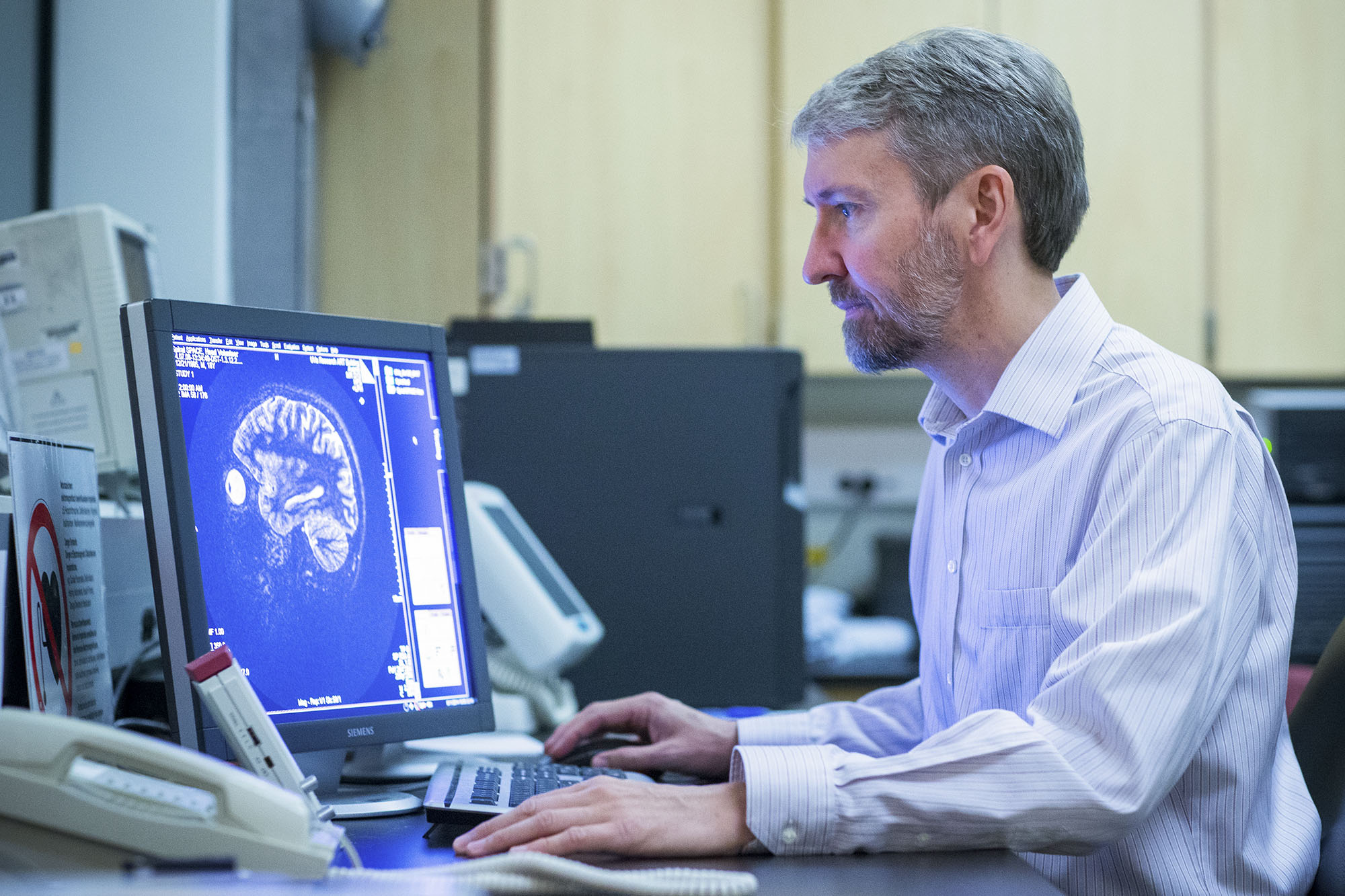
When a patient is placed inside the cramped confines of an MRI machine, the powerful magnets force many of the protons found in the water that makes up most our cells to align in the same direction, like soldiers in formation. Radio waves are then pulsed through the body, sending those protons out of equilibrium, but still held in tension by the magnets. Then, when the radiofrequency is turned off, sensors in the MRI machine analyze how fast the protons realign to the magnetic draw of the machine, and how much energy they use to do so. Knowing how long it takes different kinds of cells to realign allows the machine to paint a detailed picture of our internal anatomy.
MRIs have traditionally been instrumental in identifying diseases, like cancers and tumors, as well as in orthopedics.
Acquiring the data using the MRI machine is only part of the equation. The next big challenge is turning that data into an image that can be useful for clinicians.